
Thyroid cancer is a growing concern, with its incidence increasing globally over the years. Detecting thyroid cancer early is crucial for effective treatment, and ultrasound imaging is one of the most common tools used for diagnosis. While ultrasounds cannot provide a definitive diagnosis of thyroid cancer, they offer critical visual clues that help doctors identify suspicious nodules. One of the main questions many patients have is, “What color is thyroid cancer on an ultrasound?” This article delves into the colors seen on thyroid ultrasounds and what they signify in identifying potential thyroid cancer.
What Color Is Thyroid Cancer on Ultrasound?

Thyroid ultrasounds use sound waves to create a grayscale image of the thyroid gland and surrounding structures. In addition, color Doppler imaging is often used to assess blood flow within the thyroid. Together, these methods provide valuable insights into the nature of thyroid nodules and possible indications of cancer.
Grayscale Imaging
- Hypoechoic Areas (Dark Gray to Black):
Cancerous thyroid nodules often appear as hypoechoic, meaning they reflect less sound and therefore appear darker than surrounding tissue. This darker appearance is a key indicator of a potentially malignant nodule. - Microcalcifications (Bright White):
Cancerous nodules frequently contain microcalcifications, which appear as tiny bright white spots on the ultrasound. These are deposits of calcium and are considered a strong marker for malignancy. - Irregular Margins:
Cancerous nodules often have irregular or poorly defined borders, making them stand out against the surrounding tissue. - Taller-Than-Wide Shape:
A nodule with a shape that is taller than wide on ultrasound is more likely to be cancerous. This shape indicates invasive growth into deeper tissues.
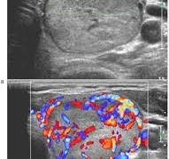
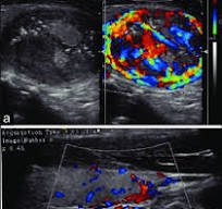
Color Doppler Imaging
Color Doppler imaging adds another dimension to thyroid ultrasound by showing blood flow within the thyroid and its nodules. The colors commonly seen in Doppler imaging are:
- Red and Blue:
These colors represent the direction of blood flow. Increased vascularity (blood flow) within a nodule, especially at the center, is a potential indicator of cancer.- Red: Indicates blood flowing toward the ultrasound probe.
- Blue: Indicates blood flowing away from the probe.
- Increased Central Vascularity:
Cancerous thyroid nodules often show increased blood flow at their center, which is visualized as a dense cluster of red and blue patterns in Doppler imaging.
Combined Indicators of Cancer
Thyroid cancer on ultrasound is not determined by one factor alone. Instead, doctors assess a combination of features, including:
- Hypoechoic appearance.
- Microcalcifications.
- Irregular margins.
- Taller-than-wide shape.
- Increased central vascularity on Doppler imaging.
Conclusion
Thyroid ultrasounds provide a wealth of information about the structure and potential abnormalities within the thyroid gland. Cancerous thyroid nodules often appear as hypoechoic (dark) with microcalcifications (bright white spots) and increased vascularity (red and blue on Doppler imaging). While these features are highly suggestive of malignancy, a definitive diagnosis requires a biopsy and further tests. If you notice any thyroid-related symptoms or concerns, consult a healthcare professional for proper evaluation and guidance. Early detection is the key to effective treatment and better outcomes.
Reference
- Yang, X., Xu, J., Sun, J., Yin, L., Guo, R., & Yan, Z. (2021). Clinical value of color Doppler ultrasound combined with serum tumor markers for the diagnosis of medullary thyroid carcinoma. Oncology Letters, 22, 561. https://doi.org/10.3892/ol.2021.12822
- Baig, Faisal & van Lunenburg, Jurgen & Liu, Shirley & Yip, Shea & Law, Helen & Ying, Michael. (2017). Computer-aided assessment of regional vascularity of thyroid nodules for prediction of malignancy. Scientific Reports. 7. 10.1038/s41598-017-14432-7.
- Singhal, Alka & Baijal, SanjaySaran & Sarin, Deepak & Arora, Sowrabh & Mithal, Ambrish & Gautam, Dheeraj & Sharma, Naman. (2018). Intrathyroidal parathyroid adenoma in primary hyperparathyroidism: Are we overdiagnosing? case series and learning outcomes. Journal of Head & Neck Physicians and Surgeons. 6. 48. 10.4103/jhnps.jhnps_38_17. source