
Lymphoma thyroid cancer is a rare type of cancer that originates in the thyroid gland. Unlike other forms of thyroid cancer, it typically arises from lymphocytes, which are part of the immune system. Detecting thyroid lymphoma early is crucial for effective treatment, and ultrasound imaging plays a significant role in diagnosis. This article explores how ultrasound is used to evaluate thyroid lymphoma and what the observed colors might indicate.
Key Takeaway:
- Thyroid lymphoma appears as a hypoechoic (dark) mass on ultrasound.
- It often has a homogeneous texture, distinguishing it from other thyroid conditions.
- Increased vascularity, shown in red and blue Doppler colors, may indicate its aggressive nature.
- Ill-defined margins are common, while calcifications are typically absent.
- Ultrasound helps identify thyroid lymphoma, but a biopsy is needed for a definitive diagnosis.
Lymphoma Thyroid Cancer Ultrasound Colors

When evaluating thyroid lymphoma on an ultrasound, doctors look for specific patterns and characteristics that differentiate it from other thyroid conditions:
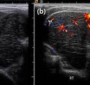
- Hypoechoic Areas (Dark Gray to Black):
- Lymphoma often appears as a hypoechoic (darker) mass, indicating that the affected area reflects less sound on the ultrasound.
- These dark regions suggest a dense, solid mass, which is typical of malignancies like lymphoma.
- Homogeneous Texture:
- Unlike other thyroid cancers that may show irregular or mixed textures, lymphoma typically appears as a uniform, homogeneous mass.
- This even texture helps distinguish it from benign nodules or other cancers.
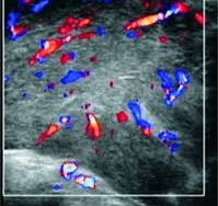
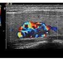
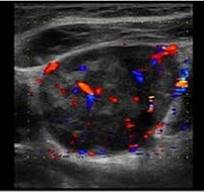
- Increased Vascularity (Color Doppler):
- Using color Doppler imaging, areas of thyroid lymphoma may show increased blood flow.
- The red and blue Doppler colors represent the movement of blood within or around the mass, indicating its aggressive nature.
- Ill-Defined Margins:
- Lymphoma in the thyroid often shows poorly defined edges, making it difficult to clearly separate the mass from surrounding tissues.
- Absence of Calcifications:
- Unlike other thyroid cancers, lymphoma usually does not exhibit microcalcifications, which would appear as bright white spots on the ultrasound.
What Do Colors Mean on a Thyroid Ultrasound?
Colors on a thyroid ultrasound, particularly with Doppler imaging, indicate blood flow:
- Red and Blue: Show the direction of blood flow within the thyroid or a nodule.
- Dark Gray to Black (Hypoechoic): Indicates solid or dense areas, often seen in malignancies.
- White (Hyperechoic): Represents calcifications or highly reflective tissues.
What Does a Cancerous Thyroid Look Like on an Ultrasound?
A cancerous thyroid nodule often has these features:
- Hypoechoic (darker) appearance compared to surrounding tissue.
- Irregular or poorly defined borders.
- Microcalcifications appear as small bright spots.
- Taller-than-wide shape, suggesting malignancy.
- Increased blood flow within the nodule on Doppler imaging.
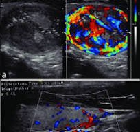
Can Thyroid Lymphoma Be Seen on Ultrasound?
Yes, thyroid lymphoma can be seen on an ultrasound. It typically appears as:
- A hypoechoic, homogeneous mass.
- A lesion with ill-defined margins.
- Increased vascularity on Doppler imaging.
What Is the Cancer Color for Thyroid Cancer?
The awareness ribbon color for thyroid cancer is teal, pink, and blue. These colors symbolize thyroid cancer awareness and advocacy.
What Are the Color Codes for Cancer?
Different cancers have unique awareness colors. For example:
- Pink: Breast cancer.
- Teal: Ovarian cancer.
- Orange: Leukemia.
- Teal, Pink, and Blue: Thyroid cancer.
What Are the Red Flags for Thyroid Cancer?
Red flags for thyroid cancer include:
- Rapid growth of a thyroid nodule.
- Hard, fixed nodules that don’t move with swallowing.
- Hoarseness or changes in voice.
- Swollen lymph nodes in the neck.
- Suspicious ultrasound features like microcalcifications and irregular borders.
What Is the Silent Warning of Thyroid Cancer?
The silent warning of thyroid cancer is a painless, slowly growing lump in the neck. Many people don’t experience symptoms until the cancer progresses, which highlights the importance of routine check-ups.
What Are the Symptoms of Lymphoma in the Thyroid?
Symptoms of thyroid lymphoma include:
- Rapidly growing neck mass.
- Difficulty swallowing or breathing.
- Hoarseness or voice changes.
- Swollen lymph nodes in the neck.
Is Thyroid Cancer 100% Curable?
Thyroid cancer has a high cure rate, especially for types like papillary and follicular thyroid cancers, which have a 5-year survival rate exceeding 98%. However, factors like cancer type, stage, and early detection impact outcomes.
Conclusion
Thyroid lymphoma presents unique patterns on ultrasound imaging, with hypoechoic regions, homogeneous textures, and increased vascularity as key indicators. While ultrasound is a valuable tool for identifying potential thyroid cancers, a definitive diagnosis often requires further tests, such as a biopsy or CT scan. If thyroid lymphoma is suspected, early evaluation and treatment are essential for better outcomes. Always consult your healthcare provider for accurate assessment and guidance.
Reference
- Castro MR, Gharib H. Thyroid nodules and cancer. When to wait and watch, when to refer. Postgrad Med. 2000 Jan;107(1):113-6, 119-20, 123-4. doi: 10.3810/pgm.2000.01.808. PMID: 10649669. https://pubmed.ncbi.nlm.nih.gov/10649669/
- Beasley MJ. Lymphoma of the thyroid and head and neck. Clin Oncol. 2012;24:345–51. doi: 10.1016/j.clon.2012.02.010. [DOI] [PubMed] [Google Scholar]
- Watanabe N, Noh JY, Narimatsu H, Takeuchi K, Yamaguchi T, Kameyama K, Kobayashi K, Kami M, Kubo A, Kunii Y, Shimizu T, Mukasa K, Otsuka F, Miyara A, Minagawa A, Ito K, Ito K. Clinicopathological features of 171 cases of primary thyroid lymphoma: a long-term study involving 24553 patients with Hashimoto’s disease. Br J Haematol. 2011 Apr;153(2):236-43. doi: 10.1111/j.1365-2141.2011.08606.x. Epub 2011 Mar 4. PMID: 21371004. https://pubmed.ncbi.nlm.nih.gov/21371004/
- Du W, Ling W, Ma X, Jiang C, Wang J, Zhu C, et al. Contrast-enhanced ultrasound in the therapeutic assessment of diffuse large B-cell lymphoma: A case report. Oncol Lett. 2017;14:4593–8. doi: 10.3892/ol.2017.6758. [DOI] [PMC free article] [PubMed] [Google Scholar]
- Wang Z, Fu B, Xiao Y, Liao J, Xie P. Primary thyroid lymphoma has different sonographic and color Doppler features compared to nodular goiter. J Ultrasound Med. 2015;34:317–23. doi: 10.7863/ultra.34.2.317. [DOI] [PubMed] [Google Scholar]
- Nam M, Shin JH, Han BK, Ko EY, Ko ES, Hahn SY, et al. Thyroid Lymphoma: correlation of radiologic and pathologic features. J Ultrasound Med. 2012;31:589–94. doi: 10.7863/jum.2012.31.4.589. [DOI] [PubMed] [Google Scholar]
- Takashima S, Morimoto S, Ikezoe J, Arisawa J, Hamada S, Ikeda H, Masaki N, Kozuka T, Matsuzuka F. Primary thyroid lymphoma: comparison of CT and US assessment. Radiology. 1989 May;171(2):439-43. doi: 10.1148/radiology.171.2.2649921. PMID: 2649921. https://pubmed.ncbi.nlm.nih.gov/2649921/
- Xia Y, Wang L, Jiang Y, Dai Q, Li X, Li W. Sonographic appearance of primary thyroid lymphoma-preliminary experience. PloS one. 2014;9:e114080. doi: 10.1371/journal.pone.0114080. [DOI] [PMC free article] [PubMed] [Google Scholar]
- Stein SA, Wartofsky L. Primary thyroid lymphoma: A clinical review. J Clin Endocrinol Metab. 2013;98:3131–8. doi: 10.1210/jc.2013-1428. [DOI] [PubMed] [Google Scholar]
- Ha EJ, Baek JH, Lee JH, Kim JK, Song DE, Kim WB, et al. Core needle biopsy could reduce diagnostic surgery in patients with anaplastic thyroid cancer or thyroid lymphoma. Eur Radiol. 2016;26:1031–6. doi: 10.1007/s00330-015-3921-y. [DOI] [PubMed] [Google Scholar]
- Bernardi S, Michelli A, Bonazza D, Calabrò V, Zanconati F, Pozzato G, et al. Usefulness of core needle biopsy for the diagnosis of thyroid Burkitt’s lymphoma: A case report and review of the literature. BMC Endocr Disord. 2018;18:86. doi: 10.1186/s12902-018-0312-9. [DOI] [PMC free article] [PubMed] [Google Scholar]
- Wu SY, Chu CH, Duh QY, Hsieh CB, Yu JC, Shih ML. Management for primary thyroid lymphoma: Experience from a single tertiary care centre in Taiwan. Formos J Surg. 2016;49:201–7. [Google Scholar]
- Pavlidis ET, Pavlidis TE. A review of primary thyroid lymphoma: Molecular factors, diagnosis and management. J Invest Surg. 2019;32:137–42. doi: 10.1080/08941939.2017.1383536. [DOI] [PubMed] [Google Scholar]
- Nam M, Shin JH, Han BK, Ko EY, Ko ES, Hahn SY, Chung JH, Oh YL. Thyroid lymphoma: correlation of radiologic and pathologic features. J Ultrasound Med. 2012 Apr;31(4):589-94. doi: 10.7863/jum.2012.31.4.589. PMID: 22441916. https://pubmed.ncbi.nlm.nih.gov/22441916/
- Naswa N, Sharma P, Nazar AH, Mohapatra TK, Bal C, Kumar R. (18) F-FDG PET/CT for initial assessment and response monitoring in a case of high grade primary lymphoma of the thyroid gland: A case report and review of literature. Indian J Nucl Med. 2014;29:94–6. doi: 10.4103/0972-3919.130291. [DOI] [PMC free article] [PubMed] [Google Scholar]